Positron Emission Tomography (PET) is a leading imaging technique used in cancer detection, neurological studies, and heart diagnostics. PET doesn’t just show organ structure—it reveals abnormal metabolic activity, often before physical changes appear.
Here’s how it works: a small amount of radioactive tracer is injected into the body, where it accumulates in metabolically active tissues like cancer cells. As the tracer decays, it emits positrons that collide with electrons, producing pairs of gamma photons (511 keV) that travel in opposite directions. A PET scanner detects these photons to create high-resolution images of the body’s internal processes.
3DπPET, Redefining PET Imaging with Dark Matter Technology
3DπPET brings breakthroughs from dark matter research into medical imaging, offering faster scans, lower radiation doses, and sharper images. Built on cutting-edge cryogenic detector technology, it represents the next generation of Positron Emission Tomography (PET). These include innovations in cryogenic photosensor technology, liquid argon (LAr) detectors, and low-radioactivity argon procurement. The DarkSide collaboration has successfully applied these technologies in physics experiments, and 3Dπ leverages them to transform PET imaging.
3Dπ shifts away from traditional segmented detectors by using a monolithic, xenon-doped LAr scintillator, read out by multiple fast cryogenic silicon photomultipliers (SiPMs) simultaneously. This unique design provides total-body (TB) coverage, significantly improves time-of-flight (TOF) resolution, reduces patient radiation doses, and allows for much faster scans. The system’s foundation in advanced cryogenic and low-background techniques borrowed from dark matter research enables higher image resolution and better detection efficiency, pushing the limits of conventional PET technology.
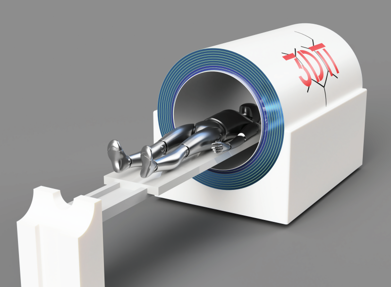
CAD model of the 3Dπ detector with a human
phantom. The ends of the cylinder are left open without a
full cryostatic enclosure to show the LAr layers.
Key features
LAr as a Scintillator: Liquid argon offers fast scintillation properties, producing more photons per unit of energy than conventional materials. Its scalability and lower costs make it ideal for large-volume detectors.
Xenon Doping: Adding xenon to LAr improves light detection and shortens the long decay time of scintillation light. This helps in clearer image formation and reduces the risk of overlapping photon signals, which could degrade image quality.
Fast Cryogenic SiPMs: The advanced SiPMs used in 3Dπ provide high photon detection efficiency (PDE) and excellent timing resolution. These sensors work well at cryogenic temperatures, making them perfect for use with LAr-based detectors, and enhance image quality through improved light yield.
How 3DπPET Stands Out?
Monolithic Detector: Captures single Compton scatter events more precisely than segmented crystals.
Reduced Scan Time: Ultra-fast electronics and SiPMs speed up the process, improving patient comfort and clinical workflow.
Lower Radiation Doses: Optimized for smaller amounts of radioactive tracer, making scans safer.
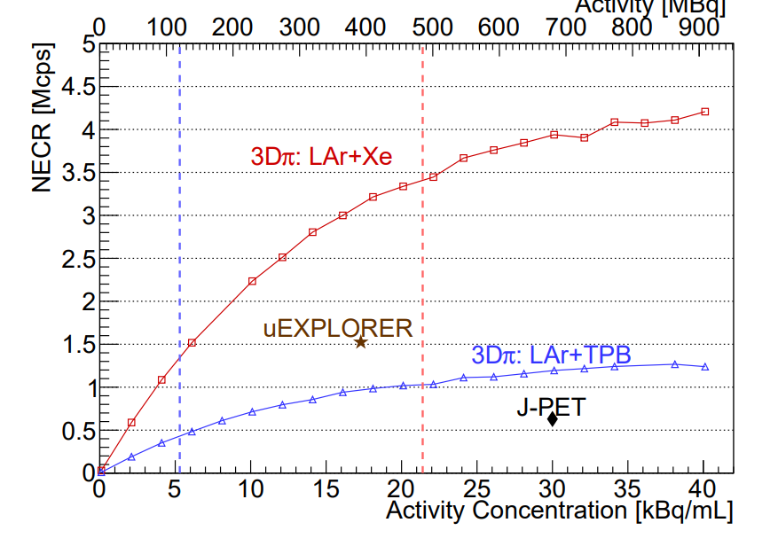
Relationship between NECR and activity
concentration for both configurations, compared with the
peak Noise Equivalent Count Rate values of uExplorer [24] and J-PET
Our Research Roadmap
The next step in our research is to secure funding for experimental work. We are actively pursuing grants to transition from simulation to building a prototype 3Dπ scanner. This experimental phase will validate the results obtained from our simulations and pave the way for real-world applications of the system in clinical PET imaging.